Science images

Mainly images of things I've taken that I think are interesting. Comments will be posted to explain what the (mainly) scientific images are. All images copyrighted by Gordon Vrdoljak. These are lower resolution images. I have higher resolution images available upon request. Send me an email specifying its intended use.

Name:
Location: Berkeley, California, United States

Friday, January 28, 2005

Wasp Head, stereomicroscope micrograph


This is the head of a wasp taken from storage in 70% ethanol, air dried, and then photographed through a stereomicroscope using a digital camera. Although not a high magnification image, you can start to resolve the compound eyes.

Thursday, January 20, 2005

sea squirt


This is from the intestine of a sea squirt.

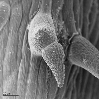
grape leaf


This is a strange looking trichome on a grape leaf.

Friday, January 07, 2005

bacteria colony on agar


This is a colony of bacteria isolated from a mixed waste sludge and then grown onto agar media. You can see a lot of attachment structures between the bacteria and agar. A number of the bacteria are dividing as well.

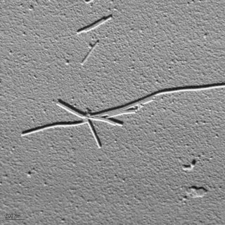
tobacco mosaic virus


Here is a scanning electron micrograph of a group of tobacco mosaic virus particles. The protein coat or capsid of the virus can be clearly seen. These were simply dried from an infected plant on silicon wafer and then coated with 1 nm of platinum followed by 10nm of carbon. Imaging with a backscatter detector allowed high resolution imaging of only the platinum and gives the cool shadow effect.

Thursday, January 06, 2005

biofilm2


Another image of the biofilm from the creek. This one is centered on a colony of diatoms in the biofilm.

biofilm


This is an image of a biofilm (jello like mush), from a creekbed. You can see diatoms (probably diatoma or fragilariaceae), the biofilm gel, and a few fungi in the image.